MRI of the brain: when necessary, how it goes, contraindications


MRI in the brain is a procedure for examining its structure without unsettling the functioning from the organ. It's employed to examine bloodstream and soft tissues to spot possible injuries and lesions as a result of strokes.
MRI is entirely safe for humans, you can find practically no contraindications. The only real limitations are related to the provision of pacemakers and metal implants. An energetic magnetic field can heat metals by the body processes or disrupt electronic mechanisms.
Indications for your procedure
MRI from the brain is needed if:
Frequent and severe headaches that cannot be addressed with medication.
Dizziness and fainting.
Numbness from the legs and arms, the look of weakness from the limbs.
A clear, crisp deterioration in memory.
Regular tinnitus.
Loss in coordination and disorientation in space.
Head trauma.
Progress
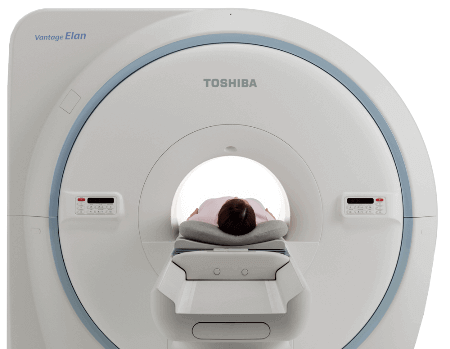
The MRI machine is really a large cylinder in which a individual is put into a supine position. Before the procedure, metal jewelry on your body, braces, as well as other metal objects are removed. The sufferer is secured with straps up for grabs to minimize mobility for accurate results.
Special products are linked to the head that generate and receive radio waves. There's significant noise inside the device, and so the patient emerged earplugs for best comfort.
Investigation result
In the resulting image, you can observe veins, neoplasms, dense and soft tissues. Picture is drawn in several projections in the desired depth, due to which the doctor will be able to assess the health of the area of the brain. In the scanning process, some images are obtained, which shows a layer-by-layer part of the cerebral tissue. Thanks to the different contrast the exact same image, every piece of information can be appreciated.
The images show: white matter, cerebral aqueduct, cerebellum, trunk, vascular structures. The tomograph creates images that are presented available as highlighted and eye shadows.
Decryption
When decrypting, a special interpretation protocol is utilized. The pictures obtained are in contrast to reference MRI data obtained from a normal brain. To accurately decipher the information received, the specialist has to thoroughly know the physiological and pathological anatomy. It can be obligatory to know the peculiarities from the operation of the tomograph, that has been employed for the examination.
Check out about glavnoe.ua explore the best net page